Canine Mast Cell Tumours

Incidence
Mast cell tumours (MCT) in dogs are very common, accounting for approximately 20% of all skin tumours in dogs. For most dogs, the underlying cause promoting the development of the tumour is not known.
Mast cell tumors can arise from any skin site on the body, and can have a variety of appearances. Veterinary oncologists recommend that before any skin lump is removed, the cells from the mass be collected for examination to rule out the lump as a mast cell (or other malignant) tumor. Mast cells are something that are easily identified on aspiration.
What you might see/ Clinical presentation
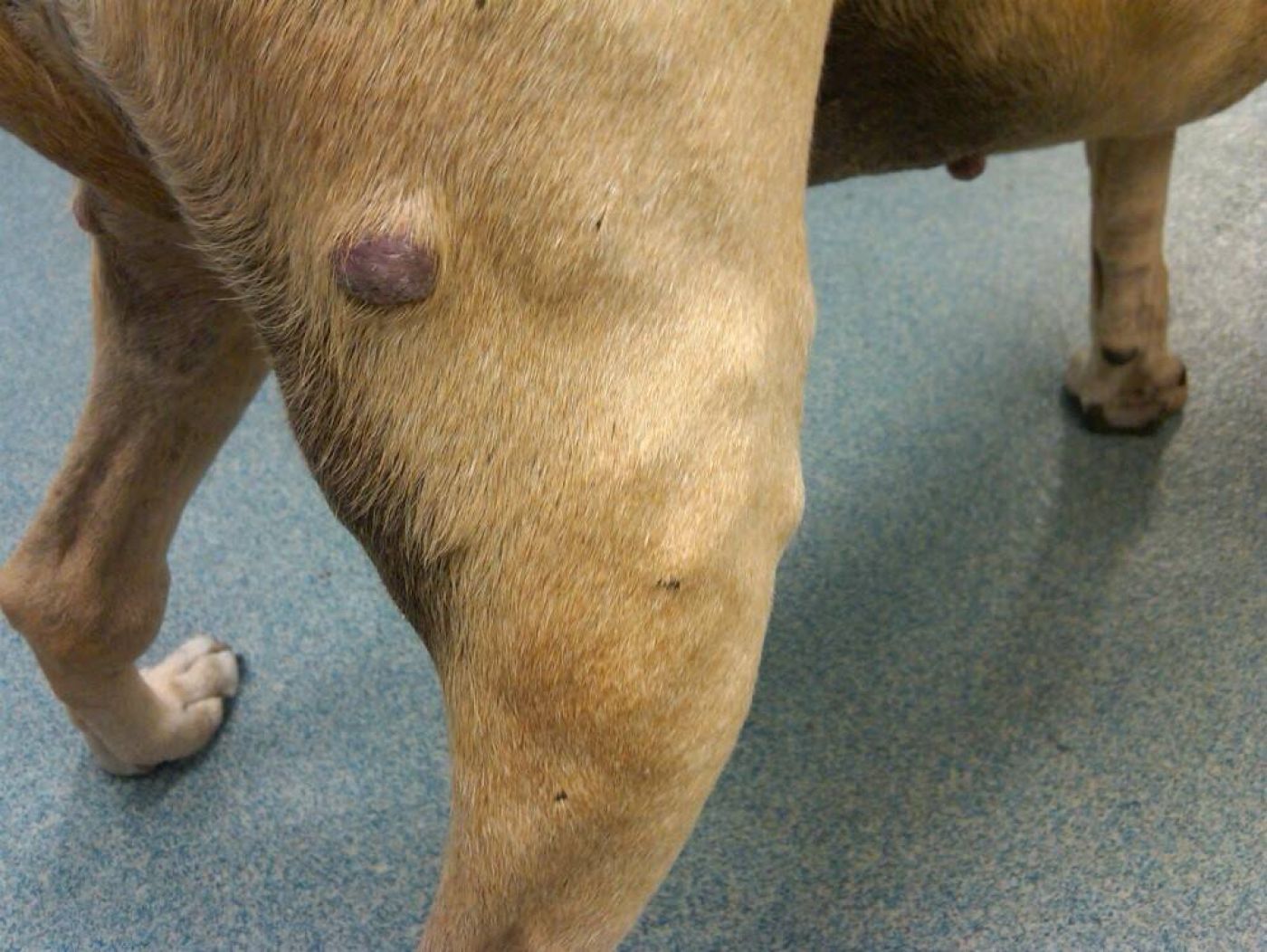
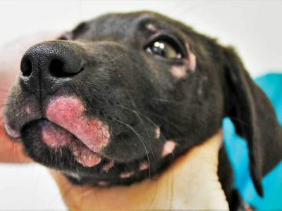
MCT most commonly are seen as solitary lumps or masses in or underneath the skin; occasional dogs can have multiple masses from MCT. MCT can look like just about anything, ranging from benign-appearing lumps (such as a lipoma), to more angry or ulcerated lumps, masses with a stalk or focal thickenings in the skin. MCT may change quickly in size (become larger then smaller ) because of reactions around the mass. Some dogs may have signs of systemic disease, which can be caused by some of the biologically active compounds found within mast cells. In most cases, evidence of a MCT is easily generated by examination of a fine-needle aspirate of the suspect mass, and aspiration is advised before removal of a mass to be sure it is not a MCT (or other skin malignancy), a finding that would demand a more aggressive surgical removal. Often, obtaining blood for a complete blood count and biochemical profile, and a urinalysis will be advised as these can help assess overall health and provide information that potentially influences treatment recommendations.
Biological behavior of mast cell tumors
Most mast cell tumors are considered locally invasive, and can be difficult to remove completely because of the extent of local spread. The behavior of mast cell tumors reflects their grade (a term used by pathologists and oncologists to describe such things as how-well differentiated a tumor is, how frequently it is dividing, how invasive to adjacent structures, and other criteria).
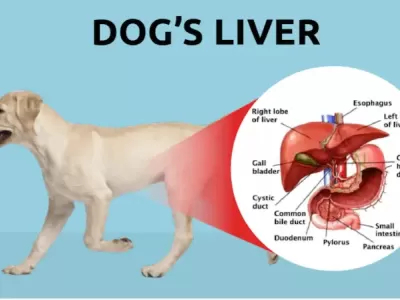
Mast cell tumors have 3 grades, with grade I being the least aggressive and least likely to spread to other organs (metastasize), and grade III being highly aggressive tumors with a high likelihood of metastasis; most grade II tumors tend not to metastasize, although they can do so. Mast cell tumors show a predilection to spread to regional lymph nodes, liver, spleen, and bone marrow.
Clinical staging (determination of the extent of the tumor)
Because of the organs to which these tumors like to spread (metastasize) to, staging a dog with a mast cell tumor (usually reserved for occasional grade I tumors, most grade II tumors and all grade III tumors) entails collection of cells from regional lymph nodes for microscopic examination, imaging the thorax (radiographs) and abdomen (radiographs, abdominal ultrasound) for enlargement of lymph nodes, liver or spleen, and some assessment of bone marrow involvement, either a bone marrow collection for microscopic examination, or examination of the white blood cells for circulating mast cells (interpreted to mean that mast cells are in the bone marrow).
Treatment options
Surgical removal is the mainstay of treatment of canine mast cell tumors. Because of their locally invasive behavior, wide margins of what appears to be normal tissue around the tumor needs to be removed to increase the likelihood that the tumor has been completely removed. For mast cell tumors that were not, or because of location, could not be completely removed, radiation therapy is often the best treatment for residual disease, although a more aggressive second surgery is possible for some dogs. Chemotherapy is sometimes used to treat mast cell tumors, but chemotherapy is usually reserved for dogs with grade III tumors; mast cell tumors are notoriously unpredictable tumors with regards to response to chemotherapy. In addition to treatment of the tumors, some dogs will be treated with medications that tend to help fight the secondary effects of the tumor. These usually include drugs like “Prednisone”, an anti-histamine like “Zantac”, and an antacid type medication like “Carafate”.
Prognosis
The prognosis for completely removed grade I and grade II tumors is excellent. The prognosis for incompletely removed grade I and II tumors treated with radiation therapy after surgery is also excellent with approximately 90-95% of dogs having no recurrence of tumor within 3 years of receiving radiation therapy. The prognosis for dogs with grade III tumors is considered guarded as local recurrence and/or spread is likely in most dogs. If your dog is diagnosed with a grade III Mast cell tumor most likely chemotherapy will be recommended as at least part of the protocol.
Key points
Dogs that develop Mast cell tumors seem to like to develop more of them and this is not necessarily the same as metastasis. So any dog diagnosed with a mast cell tumor must be watch closely in the future for the development of new tumors. As long as tumors are caught when small, surgical removal is usually adequate for treatment. Mast cell tumors can also be very unpredictable tumors. Even grade I and II tumors can behave aggressively in terms of metastasizing and or being difficult to control locally, in any given individual dog. Statistics, while useful, can never predict how an individual dog will fare with or without specific treatment.


